
Endothelial Dysfunction
What Is Endothelial Dysfunction? (Arterial Health) – The Ultimate Risk of Risk Factors
For more than a decade endothelial dysfunction has been recognized by the medical community as the critical junction between risk factors and clinical disease. It is the earliest detectable stage of cardiovascular disease. Furthermore, it is treatable, and unlike the atherosclerotic plaque that it causes, it is even reversible.
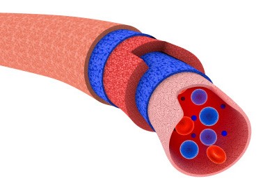
The Endothelium is the inner lining of all blood vessels, considered a “super organ” that regulates key natural biological processes that ensures homeostasis, amongst them inflammation, oxidative stress and auto-immune disease. The endothelium is the thin layer of flat, smooth cells that line the inner walls of the 62,000 miles of blood vessels in the body. As major negative lifestyle factors begin to increase, oxidative stress damages the endothelial tissue throughout the body by stiffening the veins and arteries, leading to a host of medical concerns, including erectile dysfunction, kidney disease, peripheral vascular disease, heart attack, and stroke.
Numerous studies positioned Endothelial Dysfunction as the “ultimate risk of risk factors” and is the earliest clinical detectable stage of cardiovascular disease.
Testing Against Gold Standard with EndoPAT (invasive vs, non-invasive)
These photos are of coronary arteries with excellent endothelial function (on the left) and coronary arteries with abnormal endothelial function (on the right) before and after being injected in the cath laboratory with intracoronary acetylcholine, a substance that causes the dilation of the arteries and increase in blood flow in healthy vessels.
This experimental procedure is considered the “gold standard” in assessment of coronary endothelial dysfunction and the presence of atherosclerosis in the heart.
These photos indicate the value of the EndoPAT in the assessment of endothelial dysfunction when measured against the “gold standard,” and it is this positive head-to-head assessment that earned FDA approval of EndoPAT testing in 2003.
Two photos on the left (normal endothelial function): The photo on the left shows healthy blood vessels. Following injection of acetylcholine, the blood vessels have dilated, and their increased size allows more blood flow, as noted in the right photo.
This increased blood flow is also indicated in the
before and after angiogram tracings noted directly below the photos.
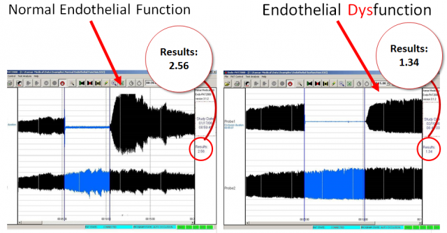
The EndoPAT results below this confirm these exact findings after blood flow was constricted in the brachial artery (left side graph) and then released (right side graph), with reactive hyperemia then measured in the fingers with EndoPAT.
Note that the red bar on the right graph, which is a marker of increased reactive hyperemia and good
blood flow, is much larger than the green bar on the left, indicating good blood flow and normal
endothelial function.
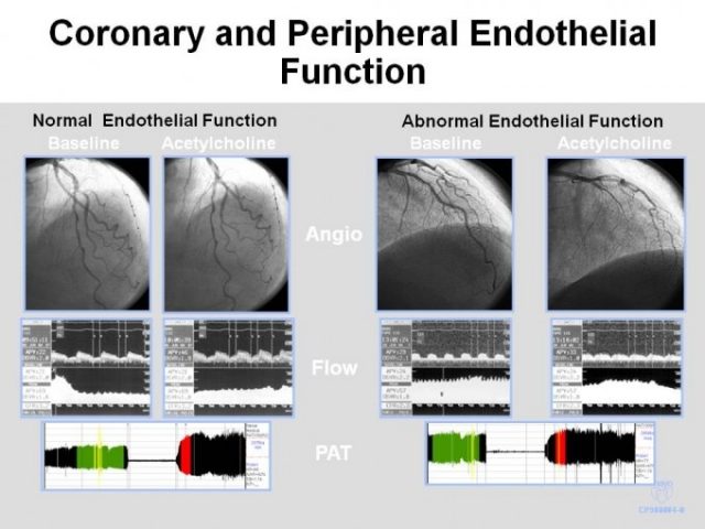
Two photos on the right (endothelial dysfunction): The upper left photo and the constricted blood vessels of the coronary artery indicate endothelial dysfunction and the presence of atherosclerosis. After injection of acetylcholine (right photo), there is further constriction of the blood vessels, since the acetylcholine went directly to the smooth muscle tissue of the damaged vessels, which increased the levels of endothelin. This caused vasoconstriction and significantly restricted blood flow in the process. Endothelin concentrations are elevated in humans with atherosclerosis. This is noted in the before and after angiogram tracings directly below the photos.
In the EndoPAT test, both the red and green bars on the left and right hand side graphs are the same size, indicating there was no reaction to the stresses created by constricting the brachial artery. The endothelium is damaged and atherosclerosis is present.
The presence of endothelial dysfunction in the coronary circulation highly predicts cardiovascular events in people in early stages of disease, beyond any conventional risk factors.
How is endothelial function measured?
In clinical cardiology research laboratories endothelial function was first measured by injecting acetylcholine via catheter into the coronary artery and monitoring the response by X ray imaging specifically measuring the diameter change of the artery. If the coronary artery diameter increases it indicates healthy endothelial function. However, if the coronary artery diameter decreases, it indicates endothelial dysfunction and a sign of atherosclerosis.
The problem with this method is that this technique is very invasive and requires hospitalization. It’s very expansive and it’s not justified for healthy or asymptomatic patients.
In 1992 scientists developed a research technique for assessing endothelial dysfunction using high resolution US imaging of the brachial artery. This technique (also called a reactive hyperemia procedure) requires 5 min occlusion and release of the brachial artery blood flow using a blood pressure cuff, and manually measuring the diameter of the brachial artery using a high-frequency ultrasound imaging probe. If the brachial artery diameter increases in less than 5% it indicates endothelial dysfunction and a sign of cardiovascular disease.
However, if the brachial artery diameter increases more than 10% it is a clear indication for healthy endothelial function. The healthier the artery – the larger the reactive hyperemic response.
Although this discovery was a major step forward and a useful tool for research laboratories it lacks feasibility in use in clinical practice and doctor offices mainly because it
requires an experienced sonographer hence its very operator dependent and sensitive to the position of the arm.
It often creates inconsistent results and unacceptable variations.
The EndoPAT ™ overcame these limitations. It features the innovative proprietary PAT® (Peripheral Arterial Tone) technology, which measures the overall health of the endothelium. The Peripheral Arterial Tonometry signal is measured from the fingertip by recording finger arterial pulsatile volume changes.
The higher the Peripheral Arterial Tonometry amplitude – the better the vascular function.
Results of the 15-minute test are automatically calculated and an EndoScore is generated, which indicates the present state of endothelial health. It is operator and Interpreter independent, providing reproducible results.