

Sleep health has an incredible impact on long-term patient wellness, and dental health is no different1. In honor of National Sleep Awareness Week, March 14-20, dentists should take time to understand what patients’ teeth can reveal about their sleep quality and their overall health.
This visual guide outlines the visible signs and common symptoms associated with obstructive sleep apnea (OSA). The American Dental Association (ADA) has identified several indicators that patients likely suffer from obstructive sleep apnea2, including:
- Bruxism
- Large or scalloped tongue
- Retrognathia
- Narrow/obstructed airway
Those suffering from OSA have a higher risk of experiencing heart disease, obesity, and diabetes, among other chronic illnesses2. As OSA is often underdiagnosed, dentists who remain educated on how to identify these visible signs will be able to advocate for and help these patients.
Learn These Four Visible Signs of Sleep Apnea
The characteristics that present the most dramatic visible indicators, their associated.
In recent years, the American Academy of Dental Sleep Medicine (AADSM) has advocated for standardization of diagnostics and treatment in dental sleep medicine3. A large part of that effort focuses on developing consistent dental education materials that show how sleep disorders like OSA may present in patients.
Practicing dentists should seek continuing education to recognize and manage their patients’ sleep disorders effectively. For OSA specifically, the characteristics that present the most dramatic visible indicators, their associated symptoms, and their typical presentations are as follows.
The characteristics that present the most dramatic visible indicators, their associated.
symptoms, and their typical presentations are as follows.
Bruxism
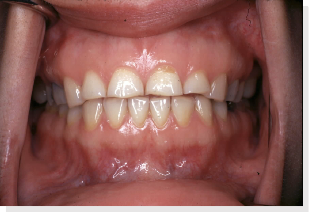
Bruxism, or teeth grinding, presents as excessive dental attrition, particularly along the incisal and occlusal surfaces of teeth.3 Unlike normal attrition, which occurs from day-to-day wear and tear, bruxism significantly damages dental crowns and exposes the fragile dentin beneath.
Over time, the mandible and maxilla bones, and therefore the upper and lower gums, come closer together as the enamel and dentin wear away and teeth take on a flattened shape3, as shown in this image (data on file). This contributes to the oral obstruction responsible for OSA symptoms.
Bruxism, or teeth grinding, presents as excessive dental attrition, particularly along the incisal and occlusal surfaces of teeth.4 Unlike normal attrition, which occurs from day-to-day wear and tear, bruxism significantly damages dental crowns and exposes the fragile dentin beneath.
Over time, the mandible and maxilla bones, and therefore the upper and lower gums, come closer together as the enamel and dentin wear away and teeth take on a flattened shape4, as shown in this image (data on file). This contributes to the oral obstruction responsible for OSA symptoms.
Over time, the mandible and maxilla bones, and therefore the upper and lower gums, come closer together as the enamel and dentin wear away and teeth take on a flattened shape3, as shown in this image (data on file). This contributes to the oral obstruction responsible for OSA symptoms.
Large or Scalloped Tongue
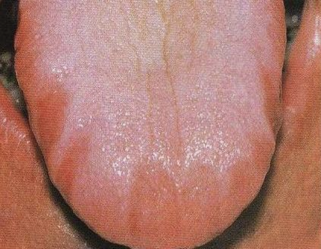
Patients with OSA often present with a scalloped tongue5, as pictured here (data on file). A scalloped tongue will appear swollen or puffy. Often, patients unconsciously push their tongues into their mandibular teeth as they strain to breathe while asleep. This leaves visible waves or notches that form uneven ridges on the perimeter of the tongue surface.
Retrognathia
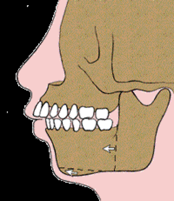
As the illustration shows (data on file), retrognathia is a specific type of malocclusion in which the maxillary teeth overhang mandibular teeth. This is also known as overclosure6, a form of teeth misalignment that is especially common among patients with bruxism. Retrognathia can worsen the vertical collapse seen in bruxism sufferers, which further constricts patient airways as seen in OSA.7
Narrow or Obstructed Airway
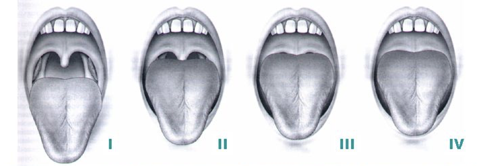
The American Dental Association describes this visible sign of OSA as “increased volume of lateral pharyngeal walls, tongue and total soft tissue.”2 As shown in these illustrations and images (data on file), obstructed airways can leave very little room for sufferers to intake full breaths of air, which is why OSA can interrupt patients’ sleep hundreds of times in a single night1.
Conclusion
These four physical characteristics are among the most dramatic visible indicators of obstructive sleep apnea found in the oral cavity.2 When dentists learn to identify these indicators and the symptoms that accompany them, patients with undiagnosed OSA are more likely to receive effective care that improves their oral health, as well as their longevity and quality of life.1
In addition to continuing education, the AADSM recommends that dentists who treat patients with OSA acquire some form of accreditation in sleep medicine3. This can include certification from a non-profit working in dental sleep medicine or an official designation based on clinical experience in the field. Doing so ensures that dentists are providing OSA patient with consistent, high-quality care that adheres to professional and ethical standards.
References:
- American Academy of Dental Sleep Medicine. Obstructive Sleep Apnea. https://www.aadsm.org/obstructive_sleep_apnea.php. Accessed March 17, 2021.
- American Dental Association. Oral Health Topics – Sleep Apnea (Obstructive). https://www.ada.org/en/member-center/oral-health-topics/sleep-apnea-obstructive. Accessed March 17, 2021.
- Levine M. Bennet K.M. Cantwell M.K. Postol K. Schwartz D.B. Dental sleep medicine standards for screening, treating, and managing adults with sleep-related breathing disorders. Journal of Dental Sleep Medicine. http://dx.doi.org/10.15331/jdsm.7030. Accessed March 24, 2021.
- James L. Clinical signs of bruxism. Dentalcare.com. https://www.dentalcare.com/en-us/professional-education/ce-courses/ce485/clinical-signs-of-bruxism. Accessed March 17, 2021.
- Holland K. What causes a scalloped tongue? Updated March 7, 2019. Healthline. https://www.healthline.com/health/scalloped-tongue
- James L. Occlusion. Dentalcare.com. https://www.dentalcare.com/en-us/professional-education/ce-courses/ce485/occlusion. Accessed March 17, 2021.
- Jenzer AC. Retrognathia. StatPearls [Internet]. Published July 7, 2020. https://www.ncbi.nlm.nih.gov/books/NBK538303/.. Accessed March 17, 2021.